Answers to problems
Chapter
2
1. A man with red-green colour blindness is
married to a girl with normal colour vision.
What is the probability that his children will be colour blind?
Answer: Red-green colour blindness is
an X-linked recessive condition. A ll
the offspring of affected males would have normal colour vision, but all their daughters would be carriers In the very rare event of his partner being
a carrier of red-green colour
blindness, half their offspring, whether male or female, would be colour blind,
half their daughters would be carriers and half their sons would be
normal.
These
results are summarized in the two following Punnett squares:
Xb
- Gene for X-linked coulor blindness; X = normal gene
1.
Affected male – genotypically normal female
|
|
X |
X |
|
|
Xb |
Xb X |
Xb X |
All daughters carriers |
|
Y |
XY |
XY |
All sons normal |
2. Affected male – carrier female (Very rare)
|
|
Xb |
X |
|
|
Xb |
Xb Xb |
Xb X |
Half daughters colour blind; half carriers |
|
Y |
Xb Y |
XY |
Half sons colour blind; half normal |
2. A boy
has Duchenne muscular dystrophy. His maternal uncle had died of muscular
dystrophy at the age of 21 years. What
is the probability that this child's sibs will be affected?
Answer: This boy has an X-lined recessive condition. The family history on the maternal side
indicates that his mother is an obligate carrier. Consequently, half his brothers would be similarly affected and
half would be normal. All his sisters
would be unaffected, but half of them would be carriers. These esults can be observed in the Punnett square below.
Xd
– gene for Duchenne Muscular dystrophy;
X – normal allele
1.
Carrier female – Normal male
|
|
Xd |
X |
|
|
X |
Xd X |
X X |
All sisters normal; half of
them carriers |
|
Y |
Xd Y |
XY |
Half his brothers affected; half normal |
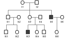 The adjacent pedigree is of a
family with haemophilia as shown in Fig.9.4 . If the gene for haemophilia is represented by Xh and
the normal allele by X', write the genotypes of each individual in the
pedigree. If the genotype cannot be determined with certainty write the
possible alternatives.
The adjacent pedigree is of a
family with haemophilia as shown in Fig.9.4 . If the gene for haemophilia is represented by Xh and
the normal allele by X', write the genotypes of each individual in the
pedigree. If the genotype cannot be determined with certainty write the
possible alternatives.
Answer:
Begin
with affected individual, III:3 . His
genotypes is Xh Y. Similarly, his affected uncle II: 5 must
have the same genotor.Because of the family history, his mother II:3 is an
obligate carrier with genotype Xh X'. His father (II:4) must be X' Y.
II:1
– X'Y (unaffected); II:2 and II:6 are
X' X'
III:1
- X'Y (unaffected); III:2 - X' X' ;
III:4
– could be either X' X' or Xh X' (carrier)
III:5
- X'Y (unaffected);
III:6
- Xh X' (obligate carrier because her father is affected)
- Explain, giving reasons,
whether the following pedigrees are compatible with autosomal dominant,
autosomal recessive or X-linked dominant and X-linked recessive
inheritance. (Note that a pedigree
may be compatible with more than one type of inheritance.)

Answers:
a.
This pedigree shows
dominant inheritance because of linear transmission from parent to
offspring. It could be either autosomal
dominant or X-linked dominant
b.
This is very similar
to the above - autosomal dominant or
X-linked dominant inheritance
c.
This pedigree is
consistent with X-linked recessive inheritance
because:
i.
only males are
affected,
ii.
transmission is
always through a carrier female, and there
is no male to male transmission
Another possibility is
autosomal dominant inheritance with decrased penetrance, thus appearing to skip
generations.
5. Examine
the pedigree from a family with a genetic disease and answer the questions
below:
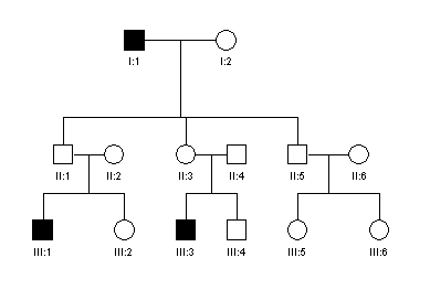
a. Does
this pedigree indicate autosomal dominant, recessive or sex-linked type of
inheritance? Give reasons for your
choice.
Answer: This pedigree is consistent with autosomal dominnat inheritance
with reduced penetrance. X-linked
inheritance is excluded because of male to male transmission (II:1 to III:1).
b. Assuming
that B and b are the normal and mutant alleles
respectively, what would be the genotypes of the individuals: II.1, II.2
and III.3 ?
Answer:
II:1 – Bb . This
individual, athough unaffected by the diease is obviously transmitting the
disease
II:2
– bb . This individual is normal and unrelated to the affected family
III:3
– Bb. Affected individual.
c.
Individual II.3 requested
genetic counselling. What is the
probability that her child would be affected. Explain why.
Answer:
II:3 is obviously transmitting the disease although she is unaffected. The chance that her children would be heterozygotes is 50%. However, only some of these would be affected, depending on the penetrance. In the aggbove pedigree, three out of five heterozygotes were affected, equal to 60%. This is only a rough measure of the penetrance because of the small number of individuals involved. Thus II:3 has a risk of 60% of 50% (=30%) that her offspring would be affected, and 20% risk that her normal offspring are in fact carriers of the abnormal gene.
6. A young
lady requested pre-marital genetic counselling because her sister had died in
infancy of gangliosidosis, an autosomal recessive disease. What is the risk
that this young lady has similarly affected offspring? What advice should be given?
Answer:
This young lady is
normal. She could be either heterozygous (Gg) carrier or normal heterozygote (GG).
Here g represents the recessive gene for gangliosidosis and G the normal
allele. Even if she is a carrier (Gg),
the chance that her husband is also a carrier would be very small, because this
is a rare disease. Hence, she has a
very small risk that her offspring would be affected.
With the availability
of genetic testing it is possible to determine whether this young lady is in
fact heterozygous or normal, and if she is heterozygous it is possible to test
her husband. If this lady is
heterozygous and her husband is normal (GG) she has no risk of having a child
with gangliosidosis.
7. A young
man requested pre-marital genetic counselling because his brother is an
achondroplastic dwarf. This condition
is inherited as an autosomal dominant disorder. What is the risk that his
offspring will be similarly affected?
What advice should be given?
Answer:
Achondroplasia
is autosomal dominant with 100% penetrance.
This young man is normal and therefore does not carry the abnormal gene
for achondroplasia. He has no increased
risk of having affected offspring. His
risks is the same as the general population risk that a new mutation will
occur, which is in fact negligibly small.
8. A young couple had their first child
affected with cystic fibrosis. What is
the risk that their future children will be similarly affected?
Answer:
Cystic fibrosis is an autosomal recessive
disorder. This young couple must both
be heterozygous carriers. The risk that
their future offspring would be affected with cystic fibrosis is 25% (1 in 4).
Chapter 3
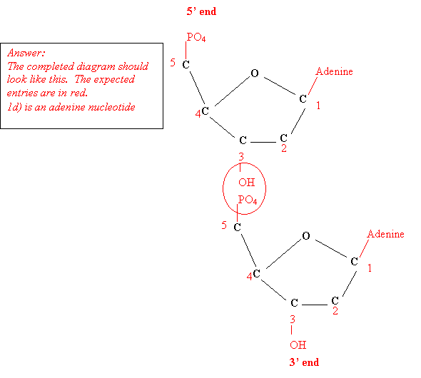
1. The diagram shows a pentose sugar.
a) Label the carbon atoms on the pentose ring.
b) Insert a PO4 group in the correct position.
c) Insert an adenine base in the correct position.
d) What is the name of the resulting nucleotide?
e) Insert a whole thymine nucleotide in the correct position to form a dinucleotide..
f) Label the 3' and 5' ends of the dinucleotide
formed.
2. The following is the sequence of bases on a single strand of DNA that was annealed with its complementary strand to form double stranded DNA.
5’ - G T T A C G G G C G G A A
C C G T A A T -3’
a) Write out the sequence of bases on the complementary strand.
3’ – C A A T G C C C G C C T T G
G C A T T A -5’
b) Calculate the proportions of the four nucleotides A,T,C and G in the single DNA strand.
Answer:
There are 20 bases in all
A =
5 bases = 25%
T
= 4 bases = 20%
C =
4 bases = 20%
G
= 7 bases = 35%
c) Calculate the proportions of the four nucleotides A,T,C and G in the double-stranded DNA formed after annealing.
Answer:
In the complementary strand of DNA there are
A =
4 bases = 20%
T
= 5 bases = 25%
C
= 7 bases = 35%
G
= 4 bases = 20%
Therefore the double stranded DNA contains
A
= 9 bases = 22.5%
T
= 9 bases =
22.5%
C
= 11 bases = 27.5%
G
= 11 bases = 27.5%
d) Compare the A:T and C:G ratios of the single and double-stranded DNA. Explain why they are different.
Answer:
The ratios of A,T,C and G are independent of one another in
single stranded DNA but T =A and C=G in the double-stranded DNA. This is because of specific base pairing in
the in double stranded DNA.
e) Work out the values of (A + G) and (T + C) for the single and double- stranded DNAs. Explain the results.
Answer:
In the given single-stranded DNA (A+ G) =12;
(T+C) = 8.
In the complementary strand of DNA (A+ G) =8 ;
(T+C) = 12.
In the double-stranded
DNA (A+
G) =20 ; (T+C) = 20.
(A+G) on one strand = (T+C) on the complementary strand.
In double-stranded DNA (A+G) = (T+C) = half the total number
of bases
3. Estimation of the proportion of bases contained in the genetic material of a virus gave the following result: Adenine 36%; Thymine 18%; Cytosine 26%; Guanine 20%. From these results determine which type of genetic material was present in this virus. Explain your result.
Answer:
In this specimen A is not equal to T and C is not equal to G. Therefore it must be single-stranded nucleic acid. Since there is thymine it must be single-stranded DNA.
A particular stretch of double-stranded DNA contianed 50 thymine and 100 cytosine bases. What is the number of adenine and guanine bases in this stretch of DNA?
Answer:
A = T = 50 bases
G = C = 100 bases
4. DNA estimations on three specimens of nucleic acid gave the following results:
A T C G U
specimen 1 20% 20% 30% 30% -
specimen 2 20% - 35% 15% 30%
specimen 3 30% 20% 30% 20% -
Identify whether each of the specimens was DNA or RNA and whether single or double stranded. Explain.
Answer:
Specimen 1: A=T and C=G. Therefore this is double stranded DNA
Specimen 2: T is replaced by U and C is not equal to
G. Therefore this is RNA
(single-stranded).
Specimen 3: Ais not
equal to T and Cis not equal to G.
Therefore, it must be single-stranded DNA.
Chapter 4
1. The following sequence is on the transcribed
strand of DNA:
3' G
C T A A T
C A G T G C G T
A 5'
Write the sequences of:
a.
the coding strand of DNA
b.
m-RNA
c.
amino acids
Answer:
a. Coding
strand
5` C
G A T T A
G T C A C G C A
T 5'
b. m-RNA
(replace the Ts in the coding strand by Us)
![]()
![]()
![]()
![]()
![]()
![]() 5` C
G A U U A
G U C A C G C A
U 5'
5` C
G A U U A
G U C A C G C A
U 5'
c. Arg Phe Val Thr His
2. The following is a schematic
diagram of DNA being transcribed:
a. DNA transcribed strand
b. DNA coding strand
c. m-RNA
The direction of transcription
is shown.
Label the 5' and 3' ends of each strand.
Answer:
Transcription always occurs in the 5’ to 3’
direction on m-RNA. Start by labelling m-RNA. The 5’ and 3’ ends
are reversed on the transcribed strand
but are the same as on the coding strand.
3. The following is the sequence
of amino acids of the last part of a protein molecule. Using the genetic
code write the codons on m-RNA and label the 3' and 5' ends. (Where more than
one codon is possible use the first one in the table.) Write the corresponding codons on the
template strand of DNA.
Answer: You must refer the table of genetic codes.
- arg -
leu - asp - phe - pro - ile -
glu - gly - val -
m-RNA 5’-AGA-CUU-GAU-UUU-CCU-AUU-GAA-GGU-GUU-3-
DNA template strand
3’-TCT-GAA-CTA-AAA-GGA-TAA-CTT-CCA-CAA- 5’
Note: do not forget to enter the
5’ and 3’ ends