Cytoarchitecture of the Cerebellum
Alfred Cuschieri
Department
of Anatomy, University of Malta
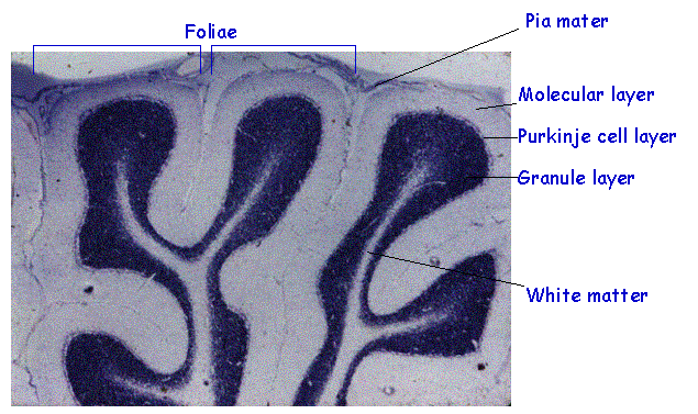
The cerebellum consists of foliae (folds) composed of cortex and white matter. The photomicrograph below is a section in which the grey matter stains blue, and the white matter is pale staining. It shows the layers forming the cerebellar cortex
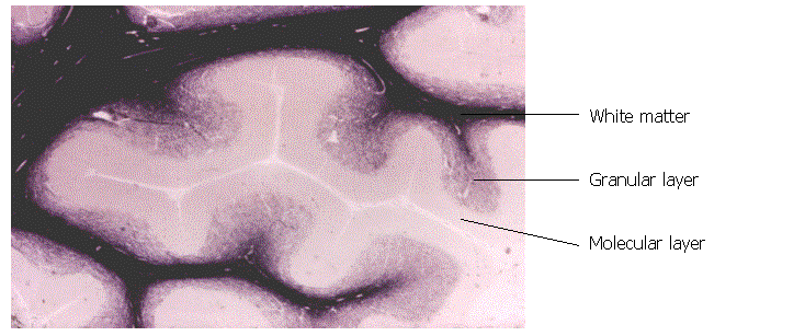
The following micrograph of the cerebellum is stained for myelin. The white matter is darkly stained. The granular layer appears as a fine meshwork of fibers. The molecular layer is pale staining
The following photomicrograph is a silver-stained section of cerebellum . It shows prominently the Purkinje cells and the neuropil in the molecular and cellular layers.
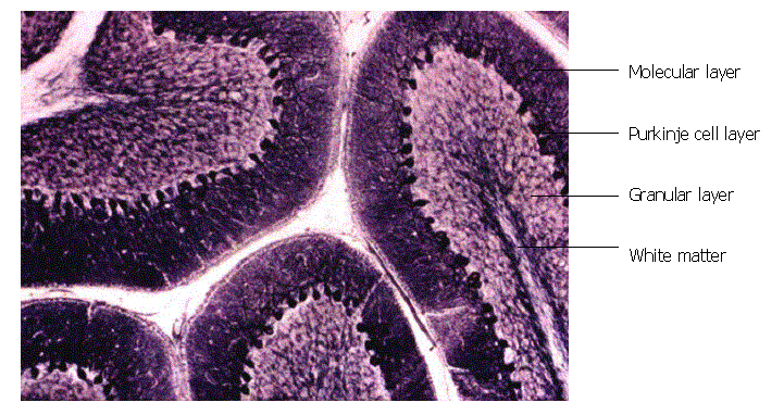
The cerebellar cortex consists of three layers on a core of white matter:
· Molecular layer
– Consists mainly of neuropil and is the site of synapses
– Contains scanty neurons consisting of stellate and basket cells
· Purkinje cell (Piriform) layer
– Single layer of neurons
– Consists of large (25 micrometer) pear-shaped neurons
· Granular layer
- Very small(7 micrometer) granular neurons
- Very numerous – 3 to 7 million neurons per cubic mm
· White matter – forms the core of the foliae
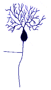
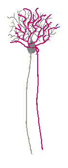
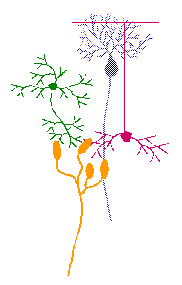
The Purkinje cells are central neurons (everything else converges on them)
 They
consist of:
They
consist of:
• A large dendritic tree in the molecular layer, which is elaborately branched and fan-shaped (branches are all in one plane)
and has dendritic spines at the sites
of synapses
• A large cell body
•
An axon which forms the efferent pathway from the
cerebellum , and sends collaterals in
the granular layer.
• GABA is the main neurotransmitter
The Purkinje cells have a definite orientation in the foliae.
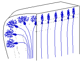
The dendritic trees of the Purkinje cells are fan-shaped in the transverse plane of the foliae.
The Purkinje cells are arranged in parallel arrays (regimentation) along the foliae
The Granule cells
• Very
numerous: 3-7 million / mm3
• Very small (7mm), closely packed neurons
• Heterochromatic nuclei, scanty cytoplasm
• Small dendritic tree in granule layer
• An unmyelinated axon
– Directed to molecular layer (centrifugal)
– Splits in T-shape manner to form parallel fiber
– Parallel fibers run longitudinally along folia
– Cross dendrites of many Purkinje cells
• Have glutamate as neurotransmitter
Note the phenomena of convergence and divergence in the cerebellum.
One granule cell diverges to contact many Purkinje cells.
About 400 parallel fibers of granule cells converge on one Purkinje cell.
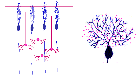
Golgi Type II Neurons Are Small Internuncial Neurons
•Situated
mostly in outer part of granular layer
•
•Dendrites extend
in 3 dimensions across blocks of Purkinje cells
•
•Short axon
with numerous arborizations in granular layer
•GABA
neurotransmitter
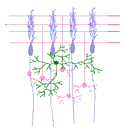
Afferents to the Cerebellum Have Two Types of Endings
1. Climbing fibers
2. Mossy fibers
They have important functional implications
Climbing fibers
•Climb through granular layer
•End in a tuft of terminal branches in molecular layer
•Correspond to dendrites of Purkinje cells (1:1 relationship)
•Are the terminal fibers of the olivo-cerebellar
neurons
•Glutamate neurotransmitter
Mossy Fibers
• Comprise
all other cerebellar afferents
• Branch and terminate in granular layer
• Have dilated endings (rosettes)
• Synapse with
– dendrites of granule cells and
– axons of Golgi type II neurons
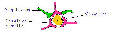
A cerebellar glomerulus is a complex of synapses having a
mossy fibre ending as its core,
synapsing with axons of Golgi type II neurons and dendrites of granule
cells
Stellate and Basket Cells Are
Situated Transversely in the Molecular Layer
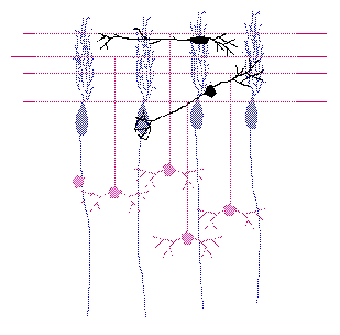
Stellate
cells
–In
superficial part, oriented transversely
–Synapse
with the Purkinje cell dendrites
–Inhibitory
Basket
cells
–In
deep part, oriented transversely
–Axons
form a basket around the Purkinje cell axon hillock
–Inhibitory
The cortico-olivo-cerebellar pathway conveys direct input from the sensori-motor and visual association areas to the cerebellum via the climbing fibers.
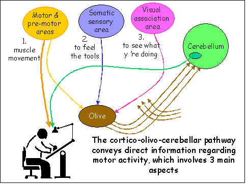
Mossy fibers convey information required for making adjustments
•
Spinocerebellar and cuneo-cerebellar
– Feedback information from muscles and joints
• Vestibulo-cerebellar
– Feedback regarding body posture & movement
• Cortico-ponto-cerebellar
– From association areas
– Motor adjustments requiring judgment, discrimination, decisions and thought
Some activities require more co-ordination than others

Cerebellar Neurons Are Stimulatory or Inhibitory on Purkinje Cells
• Climbing fibers are strongly excitatory
• Mossy fibers stimulate granule cells
• Parallel fibers of granule cells stimulate several
Purkinje cells simultaneously
• Basket cells strongly inhibit Purkinje cells
• Stellate cells inhibit Purkinje cell dendrites
• Golgi Type II cells inhibit directly the mossy fiber
input
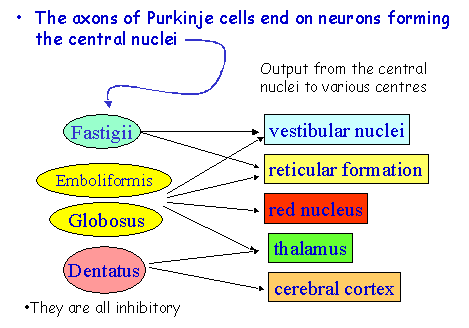
Output From the Cerebellum Is Via the Purkinje Cells
and Central Nuclei.
Signs of Spino-cerebellar Disease
• Ataxia
• Tremor
• Decomposition of movement
• Dysdiadokinesis
• Dysarthria and scanning speech
• Nystagmus
• Hypotonia
• Romberg Sign