The Fourth Week of Life:
Formation of the Embryo
Professor A. Cuschieri
Department of Anatomy
University of Malta
Objectives:
By the end of this session the student should be able to:
· Explain the mechanism underlying embryonic folding, and its role in the formation of the embryo
· Name the consequences of the formation of the head, tail and transverse folding of the embryo
· Distinguish between the skin, neural and neural crest ectoderm and their derivatives
· Explain the concept of segmentation and how it relates to the anatomical concept of myotomes
· Name the embryological origins of the vertebral bodies and vertebral arches.
· Explain how blood vessels are formed, and at which stage they develop
· Name the derivatives of ectoderm, mesoderm and endoderm
Formation of the Neural tube
 The neuro-ectoderm is
derived from the epiblast and is induced by the underlying notochord during the
third week. It is also called the neural
plate.
The neuro-ectoderm is
derived from the epiblast and is induced by the underlying notochord during the
third week. It is also called the neural
plate.
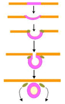
Late in the third week the neural plate begins to fold. It is first converted into a neural groove.
The neural groove deepens and eventually forms a neural tube.
Two masses of ectoderm at the edges of the neural plate, form the neural crest.
Initially the neural crest separates neuro-ectoderm from skin ectoderm.
As folding of the neural tube occurs, the neural crest cells detach from the ectoderm and form clusters that migrate into the mesoderm.
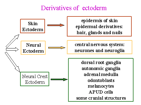
There are three derivatives of the ectoderm in this region:
1. Skin ectoderm – gives rise to the epidermis of the skin
2.  Neural crest ectoderm – are cells that migrate widely and
give rise to a large variety of structures, to be listed later.
Neural crest ectoderm – are cells that migrate widely and
give rise to a large variety of structures, to be listed later.
3. Neural ectoderm – gives rise to the central nervous system, including the neurones and neuroglia.
By the beginning of the fourth week the neural tube is formed. Closure of the neural tube begins in the region of the cervical somites and proceeds cranially and caudally. For a while the neural tube is open at both ends, the two openings being termed the cranial neuropore and the caudal neuropore.
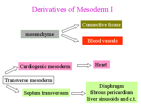
Two important masses of mesoderm
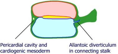
The trilaminar embryo has two important masses of mesoderm:
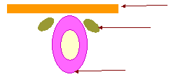
1. At the cranial end is the transverse mesoderm, in which are situated the pericardial cavity and the cardiogenic mesoderm.
2. At the caudal end is the connecting stalk that contains the allantoic diverticulum (allantoises), a small outgrowth from the roof of the yolk sac and projecting into the connecting stalk. The allantoic diverticulum will later give rise to the greater part of the urinary bladder
 Folding of the Embryo.
Folding occurs by differential growth of tissues. Neural ectoderm grows faster than the surrounding skin ectoderm and consequently fold to form a neural tube. Similarly, skin ectoderm grows faster than the underlying mesoderm and endoderm, and this differential growth causes folding of the trialminar disc and gives shape to the embryo.
Folding occurs mainly at the edges of the embryonic disc and forms three main folds:
1. Head fold
2. Tail fold
3. Lateral folds - convert the embryo into a tubular structure.
These are not three separate folds but occur simultaneously and merge into one another.
The notochord, neural tube and somites stiffen the dorsal axis of the embryo.
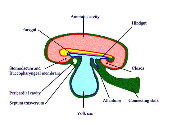
As a result of the formation of the head fold:
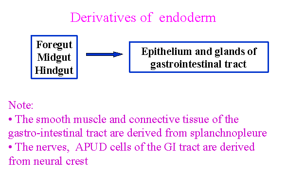
a. The foregut is formed by folding of the endoderm
b. The stomodaeum is an invagination of ectoderm, and has the buccopharyngeal membrane separating it from the foregut It opens into the amniotic cavity.
c. The pericardial cavity and cardiogenic mesoderm are shifted to the ventral aspect of the embryo and lie ventral to the foregut.
d. The part of the transverse mesoderm between the pericardial cavity and the yolk sac is the septum transversum proper. In it the liver will develop.
e. The amniotic cavity extends ventral to the cranial end of the embryo.
f. The yolk sac is constricted fro the cranial aspect.
As a result of the formation of the tail fold:
a. the hindgut is formed
b. The cloaca is an invagination of ectoderm and has the cloacal membrane separating it from the hindgut.
c. The connecting stalk is shifted ventrally
d. The allantoic diverticulum is shifted ventrally. It is an invagination of hindgut endoderm into the yolk sac.
e. The amniotic cavity extends ventral to the caudal end of the embryo.
f. The yolk sac is constricted from the caudal end
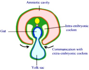
Transverse folding of the embryo
Transverse folding
a. Converts the endoderm into a primitive gut tube
b. The intra-embryonic coelom surrounds the gut tube
c. The communication between the intra- and extra- embryonic coeloms becomes constricted and eventually obliterated
Note that drastic and important changes occur in the embryonic cavities as a consequence of folding:
1. The amniotic cavity surrounds the embryo completely on all aspects and becomes the predominant cavity. It enlarges progressively.
2. The yolk sac becomes constricted on all sides, and becomes a small sac connected to the midgut by a narrow vitelline duct. It becomes progressively smaller.
3. The extra-embryonic coelom is gradually obliterated by the expanding amnion and eventually disappears completely
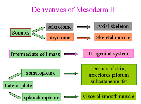
Somites
During the fourth week the embryo is segmented. Each segment consists of a somite innervated by a segmental nerve derived from a segment of the neural tube.
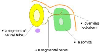
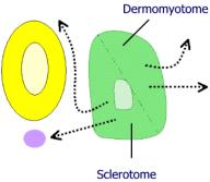
A somite is divided into two parts:
a. The sclerotome is the ventro-medial part of the somite. It contains a “cavity” of loose cells. Cells from the sclerotome migrate medially to surround the notochord and neural tube and form the axial skeleton.
b. The dermomyotome is the dorso-lateral part of the somite. Cells from the dermomyotome migrate laterally and, as its name implies, gives rise to (i) skeletal muscle and (ii) the dermis of the skin. The concept of the myotome in gross anatomy is an embryological concept. Each anatomical myotome is derived from the embryological dermomyotome that is innervated by a segmental nerve and forms a goroup of skeletal muscle cells and the dermis of the corresponding segment of ectoderm.
The neural tube induces the formation of the neural arches and their fusion across the midline.
Def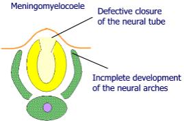 ects of closure of the neural tube will also cause failure of fusion
of the overlying neural arches. This
anomaly is termed a meningomyelocoele. As illustrated in the adjacent
diagram.
ects of closure of the neural tube will also cause failure of fusion
of the overlying neural arches. This
anomaly is termed a meningomyelocoele. As illustrated in the adjacent
diagram.
The vertebral bodies are formed from two adjacent somites
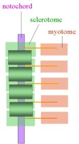
Note that the segmental spinal nerves emerge at the level of the corresponding somite, between adjacent vertebrae. The intervertebral discs correspond to the original somites and remain unossified.
Blood vessels develop throughout the mesoderm
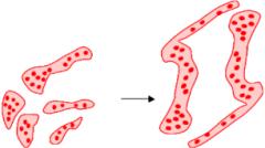 Mesodermal cells differentiate into endothelial cells surrounding a
central group of erythroblasts. These
are blood islands that coalesce to form blood vessels. Almost all parts of the
mesoderm gives rise to blood vessels.
Mesodermal cells differentiate into endothelial cells surrounding a
central group of erythroblasts. These
are blood islands that coalesce to form blood vessels. Almost all parts of the
mesoderm gives rise to blood vessels.
Differentiation of blood vessels
Blood islands and eventually blood vessels appear:
a. in the extra-embryonic mesoderm in the second week
b. in the intra-embryonic mesoderm in the third week
c. the primitve heart tube develops in the cardiogenic mesoderm (in the transverse mesoderm) at the beginning of the fourth week and a primitive circulation is established
Three structures develop in the
transverse mesoderm:
a. cardiogenic mesoderm – in which the primitive heart tubes form
b. pericardial cavity into which the heart tubes invaginate
c. the septum transversum forms part of the diaphragm, fibrous pericardium and connective tissue of the liver.
 Note that the cardiogenic
mesoderm and septum transversum are situated in the cervical region and so are
innervated from cervical segmental nerves.
Note that the cardiogenic
mesoderm and septum transversum are situated in the cervical region and so are
innervated from cervical segmental nerves.