Development of the Heart and Cardiovascular System
Professor Alfred Cuschieri, Department of Anatomy,
University of Malta
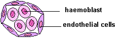 The cardiovascular system begins to develop in the third week of
gestation. Blood islands (angiocysts) develop in the newly formed mesoderm, and consist of (a) a central group of haemoblasts, the embryonic precursors of blood cells; (b) endothelial cells.
The cardiovascular system begins to develop in the third week of
gestation. Blood islands (angiocysts) develop in the newly formed mesoderm, and consist of (a) a central group of haemoblasts, the embryonic precursors of blood cells; (b) endothelial cells.
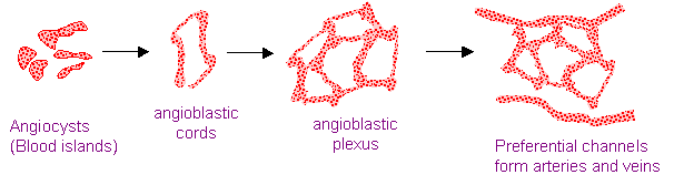
Blood islands coalesce to form a vascular plexus. Preferential channels form arteries and
veins .
Day 17 - Blood islands form first in the extra-embryonic
mesoderm
Day 18 - Blood islands form next in the intra-embryonic
mesoderm
Day 19 - Blood islands form in the cardiogenic
mesoderm and coalesce to form a pair of endothelial heart tubes
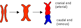
The endothelial
heart tubes fuse to form a single primitive heart tube with a
cranial (arterial) end and a caudal (venous) end.
The heart tubes are derived from the cardiogenic mesoderm situated next to
the pericardial cavity, the cranial-most end of the intra-embryonic coelom.
 Initially, at 18 days, the
cardiogenic mesoderm lies at the most cranial end of the trilaminar embryo.
Initially, at 18 days, the
cardiogenic mesoderm lies at the most cranial end of the trilaminar embryo.
After the formation of the head fold (at 20 days) the
cardiogenic mesoderm is shifted ventrally and comes to lie ventral to the
primitive pharynx.
 21 days.
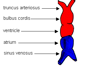
The primitive
heart tube is divided into a number of primitive chambers separated by
grooves.
The truncus arteriosus divides into a pair of
aortic arches.
The sinus venosus consists of right and left horns
 22 days.
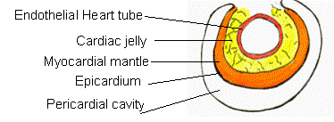
The pharyngeal endoderm induces the cardiogenic mesoderm to differentiate
into four layers, surrounded by the pericardial cavity.
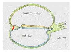
Development of a circulation
A circulation is
established during the 4th week after the myocardium is
differentiated.
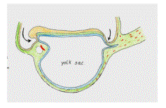
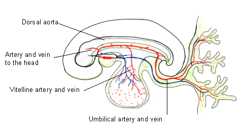
The cranial end
communicates with the paired branchial arches that open into paired dorsal
aortae. These fuse into a single dorsal
aorta. At this stage three main pairs of
arteries are present (i) to the head, (ii) vitelline arteries to the yolk sac
and (iii) paired umbilical arteries to the placenta . Three corresponding veins drain into the sinus venosus.
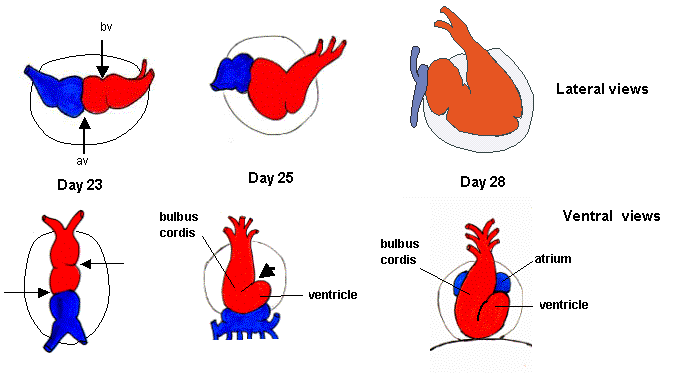
Folding of the heart tube
Folding of the heart tube occurs
on days 23-28 at two sites: (i) the bulboventricular sulcus (bv), and (ii)
the atrio-ventricular groove
(av). As a result the heart tube becomes
S-shaped.
Cardiac Asymmetry
Folding occurs
because of elongation of the heart tube, which causes it to become
asymmetrical.
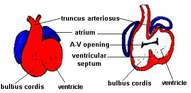 As a result of folding of the heart tube:
1.
The
atrium lies dorsal to the ventricle, bulbus cordis
and truncus arteriosus, and bulges on either side of the truncus
2.
The
bulbus cordis lies to the right of the ventricle
3.
The
ventricular septum lies between the bulbus cordis and ventricle
4.
The
A-V opening overhangs both chambers
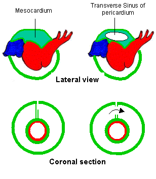 Formation of the transverse
sinus of the pericardium
Formation of the transverse
sinus of the pericardium
The heart is
suspended in the pericardial cavity by a mesocardium,
a double fold of coelomic epithelium situated in the midline.
The
mesocardium breaks down forming the
transverse sinus of the pericardium. The heart tube remains attached to the
pericardium at its cranial (arterial) and caudal (venous) ends. The transverse sinus lies dorsal to the
heart tube between the arterial and venous ends, and communicates the two sides
of the pericardial cavity. It maintains
the same relationship in the adult heart.
Development of the Sinus venosus
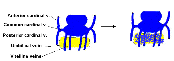 Initially the veins entering the sinus venosus are
symmetrical. During the fourth week the venous system becomes asymmetrical
causing extensive remodelling of the sinus
venosus.
Initially the veins entering the sinus venosus are
symmetrical. During the fourth week the venous system becomes asymmetrical
causing extensive remodelling of the sinus
venosus.
Initially
three sets of paired veins enter the sinus venosus:
1.
The common cardical veins enter the
sinus venosus laterally. They receive:
a.
the anterior cardinal veins from the
cranial half of the body (head, neck and upper limbs)
b.
the posterior cardinal veins from the
caudal half of the body (abdomen and lower limbs)
2.
The umbilical veins receiving
oxygenated blood from the placenta
3.
The vitelline veins drasining the
gut and yolk sac.
![]() These veins all pass through the septum transversum before
entering the sinus venosus. At the same
time the liver begins to develop within the septum transversum from cells
derived from the foregut. A venous plexus of sinusoids develops between the
liver cells and communicates with the umbilical and vitelline veins.
These veins all pass through the septum transversum before
entering the sinus venosus. At the same
time the liver begins to develop within the septum transversum from cells
derived from the foregut. A venous plexus of sinusoids develops between the
liver cells and communicates with the umbilical and vitelline veins.
Venous symmetry is radically altered by:
1.
establishment of left to right shunts in the venous
system, and
2.
obliteration of some veins draining into the sinus
venosus
Three left to right shunts are formed:
1.
A left to right
shunt between the two anterior cardinal veins. This will form the left brachiocephalic vein.
On the left, the common cardinal, the
posterior cardinal and most of the anterior cardinal veins are largely
obliterated.
2.
The ductus venosus - a preferential channel from the left umbilical to the right
vitelline veins, bypassing the liver sinusoids.
The left
umbilical vein loses its direct communication with the sinus venosus and the
right umbilical vein is obliterated
3.
The vitelline veins
communicate by three anastomoses.
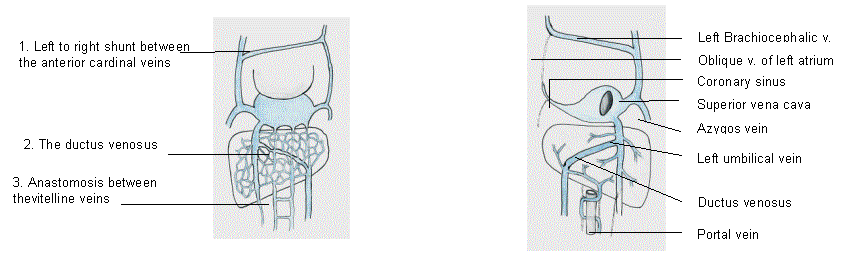
Consequences of rearrangement of the venous system
a)
The cardinal veins
*
The shunt
between the two anterior cardinal veins forms the left brachiocephalic vein.
*
On the left, the common cardinal, the posterior cardinal
and most of the anterior cardinal veins are largely obliterated. Their remnants form the oblique vein of
the left atrium
*
All the blood from the cardinal veins now drains into
the right horn of the sinus venosus
b)
The umbilical veins
*
The left
umbilical vein drains into the right
horn of the sinus venosus via the ductus
venosus, and loses its direct communication with the sinus venosus
*
The right umbilical vein is obliterated
c)
The vitelline veins
lose their direct communication with the sinus venosus. A preferential channel
fromed of parts of the right and left
vitelline veins and the three
anastomoses between them forms the portal
vein, which drains into the hepatic sinusoids.
d)
The right horn of the sinus venosus dilates
considerably as it receives all the veins.
It forms the sinus venarum part of the right atrium. The left horn of the sinus venosus 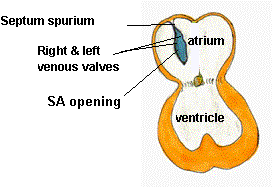 becomes small. It
forms the coronary sinus.
becomes small. It
forms the coronary sinus.
Asymmetry
of the sinus venosus shifts the sinu-atrial opening to the right of the common
atrium.
Two flaps of endothelium project into the atrium from
the sides of the SA opening forming transient right and left venous valves.
They unite near the roof of the atrium to form the septum spurium.