Development of the Heart and Cardiovascular System
II – Septation of the Heart
Professor Alfred Cuschieri, Department of Anatomy,
University of Malta
Septation
of the atrioventricular canal - Septum Intermedium.
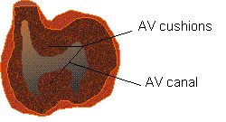 26 days :
Superior and
inferior endocardial cushions
arise from the superior and inferior walls of the atrio-ventricular canal. They
are formed of sub-endothelial thickenings of cardiac jelly.
35 days: The AV cushions fuse separating right and left AV openings.
The fused AV cushions constitute the septum intermedium
Development of the
atrioventricular valves
The
mitral and tricuspid atrioventricular valves form between the 5th
and 8th weeks. They valve
cusps and their chordae tendinae are formed by undermining of the
ventricular myocardium.
 Development of
the Atrial Septum
Development of
the Atrial Septum
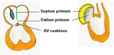
Two
atrial septa are formed, both of which contribute to the definitive atrial
septum. They are associated with two
inter-atrial communications (ostia).
1.
The septum primum begins to develop at 28 days. It is a thin, crescentic fold of endocardium that arises
craniodorsally and grows down to the AV cushions, leaving an ostium primum below its free edge. It fuses with the AV cushions at approx. 35
days, obliterating the ostium primum.
2.
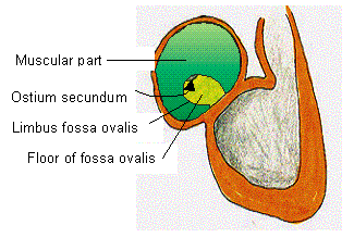
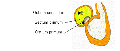 The ostium secundum
is an opening in the upper part of the septum primum. It forms at about 33 days i.e. before the ostium primum
closes. It forms by apoptosis (programmed cell death) as a
number of small perforations that
coalesce.
The ostium secundum
is an opening in the upper part of the septum primum. It forms at about 33 days i.e. before the ostium primum
closes. It forms by apoptosis (programmed cell death) as a
number of small perforations that
coalesce.
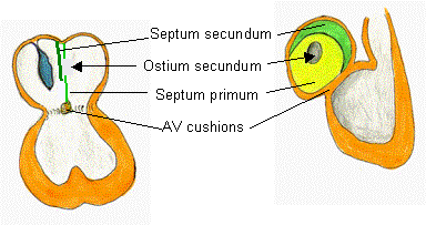
3.
The septum secundum begins to develop at about 33 days. It is a thick muscular septum that arises to the right of the septum
primum in the intersepto-valvular space (between the septum primum and the left
venous valve of the SA opening). It
grows from the roof of the atrium but never reaches the AV cushion forming
the fossa ovalis.
The
final atrial septum is formed from both septum primum and septum secundum:
The
final atrial septum is formed from both septum primum and septum secundum:
1.
The muscular part of the atrial septum is
derived from the septum secundum fused with the septum primum
2.
 The ostium secundum
is covered by the septum secundum
The ostium secundum
is covered by the septum secundum
3.
The limbus fossae ovalis is the free border
of the septum secundum
4.
The floor of the fossa ovalis is formed of
septum primum - it is thin and membranous and forms the flap valve mechanism
The Definitive Atria
The original sinu-atrial opening
communicates entirely with the right atrium.
The SA opening dilates greatly, and the right horn of the sinus venosus
is absorbed into the right atrium.
The
definitive right atrium is formed
from two parts:
a)
the muscular part derived from the
embryonic atrium - this part has musculi pectinati;
b)
the smooth part derived from the sinus
venosus (also called the sinus venarum part).
c)
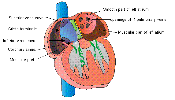
The smooth part of the
right atrium receives three openings (I) the superior vena cava; (ii) the
inferior vena cava and (iii)the opening of the coronary sinus.
d)
The crista terminalis, separating the
smooth and muscular parts, and the valves
of the inferior vena cava and coronary sinus are derived from the right
venous valve. They form one continuous curved line.
The
definitive left atrium receives no
contribution from the sinus venosus. Right and left pulmonary veins establish
communication with the left atrium. The left atrium also consists of two parts:
a)
The smooth part is derived from the pulmonary veins that have been resorbed
into the the left atrium till the level of their division. Thus this part receives the openings of the
four pulmonary veins.
b)
The muscular part of the left atrium is
derived from the left half of the embryonic atrium
Formation of the ventricular
septum
Initially, the ventricle communicates
with the atrium and the bulbus cordis communicates with the truncus arteriosus.
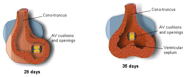
After
folding of the heart at 28 days, the bulbus cordis and truncus arteriosus are
situated to the right of the ventricle. The part of the bulbus cordis that
tapers to merge with the truncus arteriosus is the conus cordis. The conus cordis and truncus arteriosus
together form the outflow tract or cono-truncus.
 The ventricle and the bulbus cordis merge into one big
chamber. This has the AV openings
(inflow) to the left and the conotruncus (outlfow ) to the right. Two important rearrangement that occurs at
this stage are the realignment of the atrioventricular openings and the
cono-truncus to the middle of the common bulbo-ventricular cavity. This is essential for correct septation of
the ventricle.
The ventricle and the bulbus cordis merge into one big
chamber. This has the AV openings
(inflow) to the left and the conotruncus (outlfow ) to the right. Two important rearrangement that occurs at
this stage are the realignment of the atrioventricular openings and the
cono-truncus to the middle of the common bulbo-ventricular cavity. This is essential for correct septation of
the ventricle.
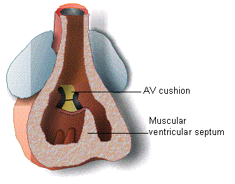
The
muscular ventricular septum grows
from the bulbo-ventricular sulcus and is directed dorsally and to the right
towards the atrioventricular cushions but does not fuse with them. Growth of the ventricular septum is arrested
at the seventh week, leaving a communication between the right and left
ventricles. This gap is closed during the eighth week by growth from
endocardial tissue, which forms the membranous part of the ventricular septum.
The
right ventricle is derived mainly from the bulbus cordis whereas the left
ventricle is derived mainly from the embryonic ventricle.
Septation of the cono-truncus
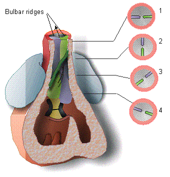
A
pair of bulbar ridges (also called conotruncal ridges) arises from
opposite sides of the cono-truncus.
They approach one another and fuse in the midline to form the spiral aortico-pulmonary septum, separating
the aorta and pulmonary trunk. The
ridges are spirally oriented, and the relative positions of the aorta and
pulmonary trunk are also spirally arranged.
The
development of the bulbar ridges begins at the lower end of the truncus
arteriosus (level 3 in Figure) and extends cranially into the truncus and
caudally into the conus. The uppermost
part of the septum fuses with the dorsal
wall of the truncus just beyond the
origin of the 6th aortic arch.
The
spiral extension of the bulbar ridges downwards into the conus forms the membranous part of the the ventricular
septum, together with a contribution from the AV cushions. This downward extension continues the spiral
and brings the aortico-pulmonary septum in line with the ventricular septum.
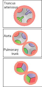 Development of Aortic and pulmonary valves
These
develop at the lower end of the truncus arteriosus. At this level there are four swellings of sub-endocardial tissue
- the right and left bulbar swellings
and two acessory dorsal and ventral
swellings. Separation of the fused bulbar ridges forms the aortic and
pulmonary vessels each containing three swellings. Growth and excavation of the swellings results in the formation
of the semilunar valves. Formation of
the semilunar valves is complete by the end of the 9th week.
Note
the positions of the valves as in adult anatomy.
The
aorta has one posterior valve and two anterior valves, above which the right
and left coronary arteries arise. The
pulmonary trunk has one anterior and two posterior valves.
Development of the conducting system of the heart
Contraction
of the heart by myogenic activity begins at about 28 days. The conducting system of the heart (SA node, AV node, bundle of His and
Purkinje fibres) consist of specialized cardiac muscle cells. The SA node is thought to be derived from
neural crest cells and is initially situated in the wall of the sinus venous. The rest of the conduction system is thought
to be derived from cardiogenic mesoderm.