The Second week of Life:
Implantation and formation of the bilaminar
embryo
Professor
Alfred Cuschieri
Department
of Anatomy
University
of Malta
During
the first week the zygote undergoes cleavage divisions. It becomes a morula and then a blastocyst,
consisting of embryoblast and trophoblast.
The whole structure, irrespective of the stage of development is
referred to as the conceptus.
On day
4 the conceptus reaches the uterine cavity, and is at the blastocyst stage.
A blastocyst is formed by accumulation of
fluid between the blastomeres. The
cells form two distinct groups:
- The trophoblast – a single layer of cells surrounding the blastocyst
cavity. It will eventually form
the placenta and membranes.
- The embryoblast – a mass of cells situated within the trophoblast. It will give rise to the embryo.
As a result of the accumulation of fluid the blastocyst increases rapidly in size. The zona pellucida ruptures and disintegrates.
The
blastocyst has a distinct polarity:
The embryonic pole is the one where the
embryoblast is situated. The opposite pole is the abembryonic pole.
The
trophoblast is now divided into two parts:
The polar trophoblast is situated at the embryonic pole and caps the embryoblast.
The mural trophoblast lines the blastocyst cavity.
On
approximately day 5 the trophoblast differentiates into two distinct layers:
1.
 Cytotrophoblast :
–
The
inner layer
–
Consists
of a single layer of cuboidal cells
–
Is
the source of dividing cells
2.
Syncitiotrophoblast
–
The
outer layer
–
Consists
of a mass of multinucleated cytoplasm with irregular finger-like processes
–
Formed
by coalescence of cells derived from the cytotrophoblast
–
Does
not contain mitotic figures
Differentiation
of the syncitiotrophoblast begins at the embryonic pole and spreads over the
blastocyst.
The syncitiotrophoblast has two important secretory
functions:
1.
Secretion
of hydrolytic
enzymes
1.
essential
for erosion and penetration of the endometrium
2.
Secretion
of Human
Chorionic Gonadotrophin (HCG)
–
Has
properties of LH
–
Is
essential for maintenance of the corpus luteum, which enlarges to form a corpus
luteum of pregnancy
–
Is
essential for maintenance of pregnancy
–
The
embryo’s signal saying “Hi Mum! I’m
here!”
Day 7
The embryoblast differentiates into two germ
layers to fom a bilaminar embryo (or germ disc):
1.
Epiblast – a columnar epithelium
adjacent to the trophoblast
2.
Hypoblast – a cubical epithelium adjacent
to the blastocyst cavity
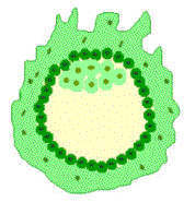
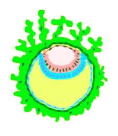
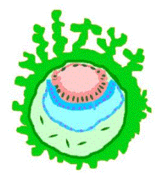
Two
cavities are also formed:
1.
Amniotic
cavity lined by the
amniotic
membrane, a thin
layer of cells derived and growing out from epiblast
2.
Primary yolk sac lined by Heuser’s membrane, a thin layer of cells derived and growing out from the hypoblast
 Separation of Heuser’s membrane from the cytotrophoblast gives rise
to two new cavities:
Separation of Heuser’s membrane from the cytotrophoblast gives rise
to two new cavities:
1.
The
secondary (definitive)
yolk sac shrinks
away from the cytotrophoblast and becomes re-lined by a new layer of cells
derived from the hypoblast
2.
The
chorionic cavity
or extra-embryonic coelom forms between the lining of the yolk sac and the cytotrophoblast.
It contains cells – the extra-embryonic mesoderm- of uncertain origin.
The chorionic cavity expands
greatly by
accumulation of fluid within it. As a result :
–
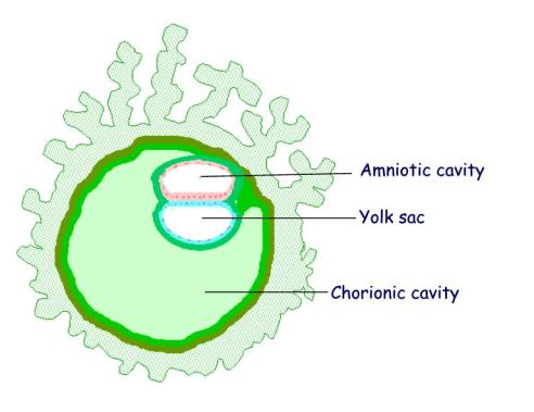 The chorionic cavity becomes the
dominant cavity
The chorionic cavity becomes the
dominant cavity
–
the
amniotic cavity and yolk sac become progressively smaller.
–
The
chorionic cavity is lined by extra-embryonic mesoderm
–
The
cytotrophoblast is lined by a layer of extra-embryonic mesoderm.
–
The
chorion is the three-layered membrane surrounding the chorionic cavity. It consists of syncytiotrophoblast, cytotrophoblast and extra-embryonic mesoderm.
–
Both
the amnion and the yolk sac are covered
externally by a layer of extra-embryonic mesoderm.
The
embryo is now a bilaminar
disc consisting of
the:
1.
 Epiblast that forms the floor of the
amniotic cavity
Epiblast that forms the floor of the
amniotic cavity
2.
Hypoblast that forms the roof of the yolk
sac
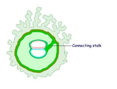
Day 14
The
embryonic disc with its amnion and yolk sac become suspended in the chorionic
cavity by a thick layer of mesoderm, which elongates to form the connecting stalk
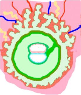 Implantation begins on approximately day 5and
is completed by the end of the second week (approximately day 13).
Implantation begins on approximately day 5and
is completed by the end of the second week (approximately day 13).
Outgrowths
from the syncitiotrophoblast invade the deciduas
They
erode the maternal blood vessels.
They
become surrounded by trophoblastic lacunae containing maternal blood
A utero-placental circulation is established by day 13
Three
stages of development of chorionic villi:
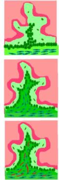
- Primary
villi
–
consist
of syncitiotrophoblast and cytotrophoblast
–
day
13
- Secondary
villi
3.
have
a core of extra-embryonic mesoderm
4.
day
16
- Tertiary
villi
–
have
blood vessels in the mesoderm
–
day
21
Anomalies
of development
- Anomalies of the embryoblast
–
“Blighted ovum” is a failure of development of
the embryoblast, while the trophoblast develops. This results in spontaneous abortion of an empty gestational sac
- Anomaly of the trophoblast
–
Hydatidiform
mole is the result
of hyper-development of the trophoblast, forming swollen and vesicular
chorionic villi. These secrete an
excess of HCG. The embryo fails to develop normally. It results in spontaneous abortion. Some hypertrophied villi may remain embedded
in the deciduas; the pregnancy test remains positive with a high titre of HCG. It may also undergo malignant change causing
choriocarcinoma.
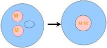
Uniparental
disomy is the
condition in which both nuclei of a zygote are derived from the same parent. Although the chromosome complement is diploid,
subsequent expression of some genes depends on whether the gene was maternally
derived or paternally derived. Thus maternal or paternal disomy give rise to
different clinical syndromes. This differential expression of genes is termed
maternal imprinting.
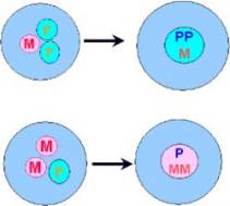
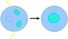 Paternal disomy occurs when both sets of
homologous chromosomes are derived from the father. This may result from dispermy or double fertilization of the
oocyte. Subsequent elimination of the maternal nucleus as a polar body results
in the zygote having two sets of paternally derived chromosomes.
Paternal disomy occurs when both sets of
homologous chromosomes are derived from the father. This may result from dispermy or double fertilization of the
oocyte. Subsequent elimination of the maternal nucleus as a polar body results
in the zygote having two sets of paternally derived chromosomes.
This
results in hydatidiform
mole.
 Maternal disomy occurs when both sets of
homologous chromosomes are derived from the mother. This may result from failure of extrusion of the male pronucleus as
the second polar body after fertilization. This results in a zygote with two
sets of maternal chromosomes.
Maternal disomy occurs when both sets of
homologous chromosomes are derived from the mother. This may result from failure of extrusion of the male pronucleus as
the second polar body after fertilization. This results in a zygote with two
sets of maternal chromosomes.
This
results in a small, underdeveloped placenta and embryo.
Paternally
imprinted chromosomes are necessary for the development of the placenta and
membranes. Maternally imprinted
chromosomes are necessary for the development of the embryoblast.
Triploidy
This
is the situation in which the zygote has a triploid rather than a diploid set
of chromosomes.
If
the extra set of chromosomes is of paternal origin, resulting from dispermy, it forms a partial hydatidiform
mole, which ends in spontaneous abortion.
If
the extra set of chromosomes is of maternal origin, resulting from failure of extrusion of the polar body, it forms the triploidy syndrome. The foetus may survive beyond the embryonic
period but ends in foetal or neonatal death.