Pattern Formation in the Embryo
Alfred Cuschieri
Department of Anatomy
University of Malta
Objectives:
o Distinguish clearly between determination, induction and differentiation
o Distinguish between transcription factors, signalling molecules and cell adhesion molecules, and give examples of each.
o Explain the importance of homeobox genes as transcription factors involved in early segmental patterning of the embryo
o Outline how BMP-4, sonic hegdehog and and activin, as examples of signalling molecules , are involved in patterning of the embryo
o State which are the periods of blastogenesis, morphogenesis and organogenesis.
Development is the formation of specialized cells, tissues and organs from a single cell, the zygote.
During development cells differentiate to produce cells of specialized function.
Differentiation is the variation in the pattern of expression of a common set of genes to form cells of diverse morphology and function.
Determination is the commitment of a cell to undergo differentiation. It is an irreversible process but is not accompanied by morphological changes.
Determinants are the cytoplasmic effector molecules that mediate determination.
Induction is the stimulation of a cell to differentiate in response to a stimulus produced by another cell. It is mediated by inducer substances that diffuse from one cell to another. It results in cell determination. Once determined cels are no longer responsive to other inducer molecules.
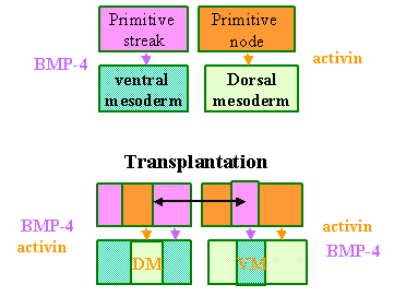 Transplantation
experiments in embryos demonstrated the processes of induction and
determination. The adjoining diagram illustrates how transplantation of a part
of the primitive streak to the primitive node and vice-versa result in the
formation of dorsal and ventral mesoderm at abnormal sites. This led to the
conclusion that the primitive streak induced the formation of ventral mesoderm
and the primitive node induced the formation of dorsal mesoderm. Further
experiments led to the discovery that BMP-4 (Bone Morphogenetic Protein– 4) and
activin were the molecular determinants
of the induction processes.
Transplantation
experiments in embryos demonstrated the processes of induction and
determination. The adjoining diagram illustrates how transplantation of a part
of the primitive streak to the primitive node and vice-versa result in the
formation of dorsal and ventral mesoderm at abnormal sites. This led to the
conclusion that the primitive streak induced the formation of ventral mesoderm
and the primitive node induced the formation of dorsal mesoderm. Further
experiments led to the discovery that BMP-4 (Bone Morphogenetic Protein– 4) and
activin were the molecular determinants
of the induction processes.
There are three classes of molecules that are important in development. The main ones are:
a. Transcription factors
b. Signalling molecules
c. Cell adhesion molecules
Transcription factors are molecules that:
a. Act in the cells that produce them
b. Bind to DNA and controls transcription of other genes in the cell
c. Initiate patterns of gene expression
Some examples of transcription factors are:
o Homeodomain or Hox genes
o Zinc finger molecules – these are steroid-binding transcription factors
o Basic helix-loop-helix proteins – such as myogenic regulatory factor
o Winged helix – such as hepatocyte nuclear factor-3.
Signalling molecules are molecules that:
a. Exert their effects on other cells
b. Mediate some of the most important interactions e.g. induction
c. Bind to transmembrane receptor molecules
d. Are mostly growth factors.
Examples are:
o Tranfsorming growth factor B (TGF-B) –activate posterior Hox genes
o Fibroblast growth factor (FGF) – activate anterior Hox genes
o Nerve growth factor (NGF) – stimulate nerve growth
o Hedgehog proteins – mediate early inductive interactions.
(The various growth factors in the above examples have many other functions apart from the main ones stated here.)
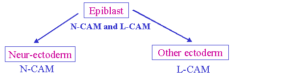 Cell adhesion molecules are:
a. Responsibe for specific cell aggregation and sorting
b. Some are Calcium-dependent molecules – Cadherins
c. Some are Calcium-independent molecules (CAMs)
An example of the function of CAMs in development is illustrated by the induction of Neuro-ectoderm and Skin-ectoderm by the differential expression of N-CAM . and L-CAM.
Developmental Field Theory.
A development field is a group of cells that differentiate in a coordinated manner.
Blastogenesis includes the developmental changes that occur from zygote formation to gastrulation in the third week.
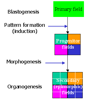 During blastogenesis
the whole embry is the primary field.
Initially the blastomeres are totipotent i.e. they can
differentiate into any cell of the body.
Recall that separation of the bilaminar embryo into two groups to form
identical twins results in the formation of two complete embryos.
During blastogenesis
the whole embry is the primary field.
Initially the blastomeres are totipotent i.e. they can
differentiate into any cell of the body.
Recall that separation of the bilaminar embryo into two groups to form
identical twins results in the formation of two complete embryos.
Pattern formation is the process of induction that leads to the formation of a number of progenitor fields, each of which is committed to differentiate into specific groups of cells – i.e. the cells become pluripotent, they have lost some of their potency, but can still differentiate into a restricted variety of cells.
Morphogenesis is the process by which the progenitor fields become secondary or epimorphic fields in which the rudiments or primordia of the future organs develop. This is the process of oganogenesis.
Three main patterns are formed during blastogenesis:
1. Establishment of the body axis
2. Formation of dorsal and ventral mesoderm
3. Segmentation.
Each of these is controlled by cascades of genes.
 Establishment of the body
axis
Establishment of the body
axis
The posterior marginal zone (caudal end of the embryonic disc) secretes activin, a signalling molecule that belongs to the TGF-B family of molecules. This induces the formation of the primitive streak, which in turn induces the formation of the cranio-caudal axis.
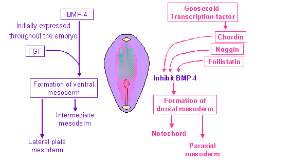
The determination of the dorsal and ventral mesoderm depends on BMP-4 (bone morphogenetic protein) and FGF (fibroblast growth factor) that are initially expressed in the whole embryo and induce the formation of ventral mesoderm, which gives rise to the intermediate mesoderm and the lateral plate mesoderm.
Expression of various transcription factors (goosecoid, chordin, noggin and follistatin) inhibit BMP-4 (and thus inhibits ventral mesoderm formation), allowing the differentiation of the dorsal mesoderm, which in turn leads to the formation of the notochord and the paraxial mesoderm.
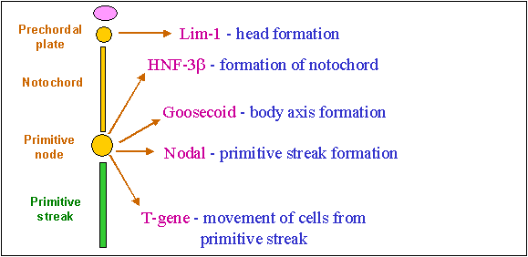
Genetic Organization Of The Primitive Streak
This diagram shows how the signalling molecules released by the primitive streak and primitive node determine the formation of other structures.
Homeotic genes
Homeotic genes are ones that control the development of segmental organization of the body structures.
They are genes that:
a. Consist of 180 bases, producing a protein consisting of 60 amino acids
b. Encode a homeodomain, a segment that binds to DNA
c. Act as transcription factors
d. Include a homeobox, an evolutionarily conserved sequence that is very similar in most organisms
e. Arose from a common ancestral gene by tandem duplication
Homeotic gene clusters
o Homeobox genes are collected into homeotic clusters.
o There are 4 homeotic clusters, labelled A,B,C and D,
o Each cluster is situated on a different chromosome.
o Each homeotic cluster consists of 13 homeotic genes numbered sequentially from 1 to 13.
o 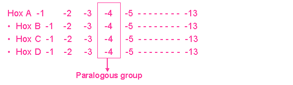 The four numerically corresponding
genes fro the four different clusters form a paralogous group. Thus there are 13 paralogous groups , as
illustrated in the diagram below.
The four numerically corresponding
genes fro the four different clusters form a paralogous group. Thus there are 13 paralogous groups , as
illustrated in the diagram below.
o The homeobox genes are responsible for patterning along the antero-posterior axis.
o The genes are expressed sequentially beginning with the paralogous group 1, which is expressed first
o The sequential genes specify different segments in cranio-caudal sequence extending from paralogous group 1, which specifies the most cranial structures, to paralogous group 13, which specifies the most caudal structures.
o Thus the first genes to be expressed specify the most cranial structures while the last to be expressed specify the most caudal structures. This is responsible for the cranio-caudal sequence of development, where the more cranial segments develop slightly before the more caudal structures. Consequently the upper limb develops ahead of the lower limb.
Establishment Of Right – Left Symmetry
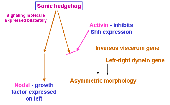 The signalling molecule
known as Sonic hedgehog (Shh) is initially expressed uniformly
throughout the embryo. It stimulates
the expression of the growth factors called nodal and lefty. Activin is expressed
unilaterally on the right, and inhibits Shh expression. Thus nodal is expressed only on the left and this results
in asymmetric development. There are
other genes that are expressed unilaterally.
These include the situs inversus gene and the left-right
dynein gene.
The signalling molecule
known as Sonic hedgehog (Shh) is initially expressed uniformly
throughout the embryo. It stimulates
the expression of the growth factors called nodal and lefty. Activin is expressed
unilaterally on the right, and inhibits Shh expression. Thus nodal is expressed only on the left and this results
in asymmetric development. There are
other genes that are expressed unilaterally.
These include the situs inversus gene and the left-right
dynein gene.
A time-table of landmarks in early human development
Day 1 - cleavage
Days 2-4 - morula; free-floating conceptus in uterine tube
Days 5-6 - formation of the blastocyst and embryoblast;
- implantation
Week 2 (days 7-14)
- formation of the bilaminar embryo 0.1 mm
Week 3 (days 15-20)
-formation of the trilaminar embryo 1.0 mm
Week 4 (days 21-28)
Day 21 - formation of neural tube 2.0 mm
Day 22 - formation of the heart
Day 23 - formation of eye and ear rudiments
Day 25 - formation of branchial arches
Day 26 - formation of upper limb bud
Day 28 - formation of the lower limb bud 5.0 mm
Weeks 5 to 9 (2nd month)
- Period of organogenesis
Week 6 1.0 cm
Week 9 4.0 cm
End of embryonic period
***********************