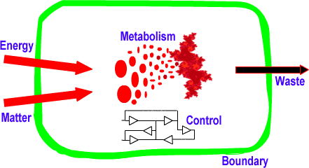
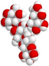
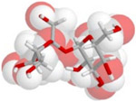
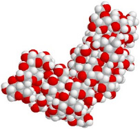
There is only one example of life we know about, that on Earth. It is based upon the cell, of size about (bacteria), made up of organic molecules in water (50-90% by mass).
Apart from water, there are, in a typical generic cell:
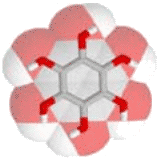
Metabolism with proteins, made up of amino acids, acting as enzymes or as structure. (50-60% of cell)
Control by genes, made up of nucleic acids (DNA), comprising the instructions. (25%)
Boundary consisting of a membrane of lipids. (10-15%)
Food storage of granules of carbohydrates. (5-15%)
Such macromolecules are sensitive to high temperature (+10°C), pH (7±0.5), and UV (>4.8eV).
They are surrounded by water concentrated with ions (K+, Na+, Cl-, Ca2+, HCO3-,...) and metabolites.
Overall, the cell consists of (more than) atoms :
The way that cells intake energy and matter differs:
Energy
Phototrophs get energy from sunlight
chemotrophs get energy from ingested reduced compounds
Matter
autotrophs get H, O, C, N, P and S from water, atmospheric CO2 and N2, nitrates, phosphates and sulphates
heterotrophs get matter from other lives (mostly).
Moreover, species may be heterotrophic for carbon but autotrophic for sulfur, say.
[For example, plants are photo-autotrophs "purple bacteria" are photo-heterotrophs methanogenic bacteria are chemo-autotrophs animals are chemo-heterotrophs]
Membrane
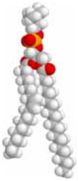
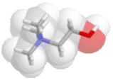
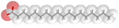
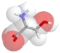
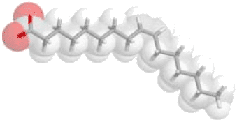
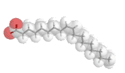
The membrane is made up of lipids, consisting of glycerol attached to two fatty acids and a 'head' molecule:
Lipid molecule"Head" lecithin+-phosphateglycerolfatty acids (16-18 C)
There are various types of "head" molecules and fatty acid "tails" that are used.
The "head" molecule is polar (+/- charges) while the "tail" is neutral; so water is attracted to the head away from the tail. The water presses the lipid molecules into a thin bi-layer surface. With the right mixture of kinked fatty acids in the lipids, the lipid bilayer is fairly flexible and naturally forms spheres (cells). If enough more lipids are added to the surface, it divides into two. The lipid molecules on the outside and inside are different.
Na+Cl-H2PO4-K+
Small neutral molecules (O2, CO2, CH3CH2OH) can diffuse through the layer; larger ones get trapped in the layer. But ions and polar molecules (H2O) are attracted to the polar heads, and rarely pass through. So the lipid bilayer is semi-permeable to soluble molecules, barely permeable to polar molecules (almost water-proof), but practically impassable to ionic molecules; it acts as a dielectric insulator separating the inside ions from the outside. It can maintain a voltage of across it with about an ion on each surface every lipid molecules. The whole cell can thus acquire about of electric energy, by pumping protons or Na+ to the outside. If ions accumulate inside the cell, water follows (by osmosis), with the risk that the lipid bilayer ruptures.
The membrane contains many proteins that regulate communication between the interior and the exterior.
Nutrients enter the cell through membrane proteins that have a hydrophilic channel. Once inside, they are attached to phophate ions (phosphorylation) to prevent the small useful molecules from diffusing back out.
Ions are actively pumped in/out by special controlled proteins. In particular, Na+ and Cl- are pumped out, while K+ and H2PO4- are pumped in. 'Aquaporin' channel proteins let in water to regulate internal concentration.
Carbohydrates
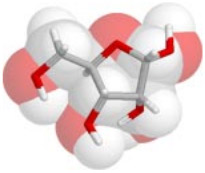
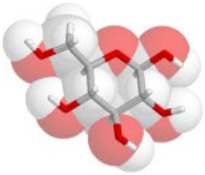
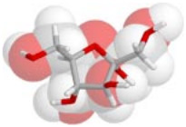
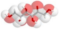
The basic building blocks are 6-sugars (hexoses) such as fructose and glucose, and 5-sugars such as ribose.
Ribose
Glucose
Fructose cyclic and acyclic forms
Each sugar molecule has various forms:
aldose or ketose forms: placement of the double-bonded O, e.g. glucose/fructose, ribose/ribulose, etc.
cyclic or linear forms: they are often interchangeable, but the linear form is more reactive; inside cells, they are mostly in the cyclic form.
isomers in the placement of the OH groups, e.g. glucose, galactose, mannose are nearly identical; there are also mirror-image isomers that are not used inside a cell.
These simple sugars can be combined together to form
sucrose (fructose+glucose), or
lactose (glucose+glucose)
They also combine to make much larger polymers, called carbohydrates, such as glycogen, starch and cellulose.
Sucrose
Small part of starch
Proteins
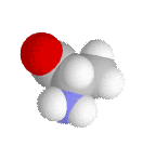
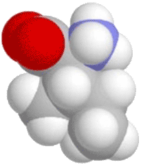
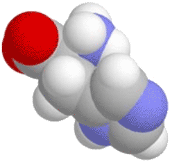
Each protein is built up as a chain of amino acids.
COO-NH3+side-chain
An amino-acid is characterised by its COO- and NH3+ functional groups, with different possible side-chains coming out of its central Carbon atom. They can join up, with the NH3+ of one amino acid reacting with the COO- of another to eject a water molecule; the joined amino acids still have these functional groups at their ends, so the process can continue to form long chains, called polypeptides.
There are at least 20 types of amino acids used by cells.
The most commonly used amino acids are neutral, and serve to make the structural bulk of the protein.
Glycine
Alanine
Leucine
Isoleucine
Valine
Proline is very rigid so it forms kinks in polypeptides
These two have an additional negatively charged COO- functional group...
Glutamate
Aspartate
...while these two have extra positively charged NH3+ functional groups:
Lysine
Arginine
These amino acids are slightly polar, and are therefore somewhat hydrophilic:
Serine
Threonine
Asparagine
Glutamine
These two have built-in cyclic structures:
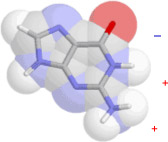
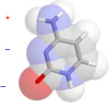
Phenylalanine
Tyrosine
The following are less used amino acids:
Methionine
Methionine, and cysteine, have a sulphur atom which can form cross-links to other amino-acids.
Cysteine
Tryptophan
Histidine
Histidine has the reactive imidazole ring.
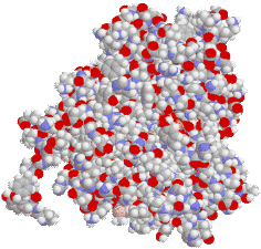
Proteins
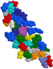
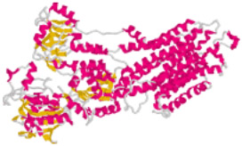
Amino acids can join with each other indefinitely. As they form a long string, they fold into a stable "secondary" structure, depending on the composition of amino acids.
[Colors are often used to distinguish amino acids, although the individual atoms are all mostly C,N,O,H.]
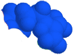
The complete chain, called a polypeptide, can have anything from hundreds to tens of thousands of amino acids (typically around 500). Their surface have regions which are hydro-phobic or hydro-philic.
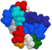
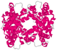
The polypeptide may be the final functional protein. But in other cases (as in the example above), whole polypeptides fit snugly into each other to form the protein, held by complementary charges along their surface. Some are meant to aggregate into larger "complexes", or even to form indefinitely long fibers.
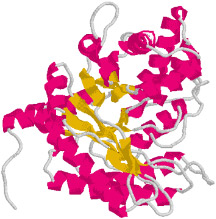
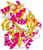
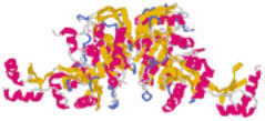
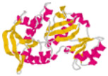
Since the underlying backbone and structure of the protein may not be apparent, it is often drawn in "cartoon form": (mouse over)
Structural motifs that appear frequently in proteins are "helices" (red ribbons) and "sheets" (yellow).
It is the shape of the protein, and the positioning of its hydrophilic amino acids, that gives it its function. The protein's function may be as a:
enzyme to catalyse reactions, or synthesize molecules,
structural element, such as fibers,
channel or transporter protein that imports/exports molecules across the membrane
regulatory protein that signals to or controls other proteins, ...,
cofactor or chaperone to temporarily store, bind, or protect other molecules
...
Enzymes act by holding on to specific molecules (substrates) at specific regions on them that match up (by hydrogen bonding). The molecule may bend slightly because of the attraction.
A nearby region in the enzyme then gives or takes an electron or proton, or a whole functional group, to the substrate, causing a change, i.e., a reaction.
Other enzymes work in the reverse fashion: two substrate molecules are "caught", and then it becomes much more likely that they react together.
Enzymes often contain a metal cation (Fe, Zn, Mn, Cu, Se,...) at their core reactive sites; and they may be physically linked in a complex with other enzymes to keep them close together and increase efficiency.
Typically about 100 molecules/second/protein react.
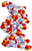
Nucleic Acids
The instructions for the control of metabolism are saved in the form of a sequence of nucleic acids.
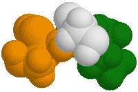
There are two pairs of complementary nucleic acids, each a purine fitting perfectly with a pyrimidine:
Guanine
Cytosine
Adenine
Thymine (or uracil)
Purines
Pyrimidines
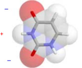
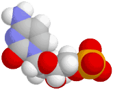
Each nucleic acid is attached to a phosphorylated ribose sugar molecule:
The OH and the phosphate groups can bond together.
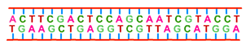
Thus they can link up together to form a long spiralling chain of nucleic acids, called DNA.
top view
side view
As more ribo-nucleic acids bond together, they fold up in the shape of a double-helix, with 10 base-pairs per turn. Notice how the helices have one wide and one narrow 'groove'.
All the information of the cell is contained in it:
DNA is quite secure because each nucleic acid molecule is paired by its complementary nucleic acid which acts both as a back-up as well as protection.
Stretches of nucleic acid sequences in DNA, called genes, code for amino-acid sequences in proteins. Roughly speaking, each gene codes for one protein. By changing the sequence of amino acids, an endless variation of proteins is made possible.