The phylum Echinodermata contains 5 classes
including the Crinoidea or feather
stars, Asteroidea or star-fish,
Ophiuroidea or brittle
stars, Echinoidea or
sea-urchinsand Holothuroidea
or sea cucumbers. These animals are relatively highly organized
animals, with their bodies constructed on a 5-rayed pattern and with a
well-developed calcareous skeleton. The spines attached to the external
skeleton can be of all sizes, from short, thick sticks to fine long
needles. They can be moved and turned in all directions by means of
muscles. The 5-rayed body is apparent in the distribution of the
sucker-like "tube-feet" in grooves called "ambulacra", separated by the
inter-ambulacra which are often spiny. The class that frequently is
related with harmful effects is the Echinoidea
or sea-urchins.
Sea-urchins are
mostly found in shallow water and along the shore, but species are also
found at depths of 4500 m. Their body is usually ball or egg-shaped,
more or less domed, rarely flattened and disc-shaped. There are two
groups of sea-urchins, those with symmetrical near-spherical bodies,
and those with usually oval bilaterally symmetrical bodies. The spines
may be long lances or short bristles, thin and with needle-points, or
thick and blunt. They break easily and may be connected with toxic
glands. Certain species of sea-urchins have venom organs (globiferous
pedicellariae) which have calcareous jaws capable of penetrating human
skin, but injuries from these are rare. Far more common are injuries by
sea-urchin spines which can break off in the skin and give rise to
local tissue reactions.
 Common injuries from sea-urchin spines usually belong to
the non-poisonous swallow water species
Arbacia lixula (Linn.), Sphaerechinus
granularis (Lam.), Psammechinus
microtuberculatus (Blainville) and Paracentrotus lividus (Lam.).
Common injuries from sea-urchin spines usually belong to
the non-poisonous swallow water species
Arbacia lixula (Linn.), Sphaerechinus
granularis (Lam.), Psammechinus
microtuberculatus (Blainville) and Paracentrotus lividus (Lam.).
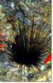 The long-spined Mediterranean species: Centrostephanus longispinus has
poisonous spines, but injuries from this species are rare since the
animal lives in relatively deep water from 10 m.
The long-spined Mediterranean species: Centrostephanus longispinus has
poisonous spines, but injuries from this species are rare since the
animal lives in relatively deep water from 10 m.
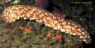 Another echinoderm species which may possibly cause
panic after handling is the sea cucumber species Holothuria forskali Della Chiaje.
When attacked or disturbed, this species extrudes long sticky whitish
threads from the hindgut, the so-called "Cuvierian tubes". These
threads contract to tighten but are easily removed. They can sometimes
cause inflammation of the skin.
Another echinoderm species which may possibly cause
panic after handling is the sea cucumber species Holothuria forskali Della Chiaje.
When attacked or disturbed, this species extrudes long sticky whitish
threads from the hindgut, the so-called "Cuvierian tubes". These
threads contract to tighten but are easily removed. They can sometimes
cause inflammation of the skin.
Many sea urchin species are edible. However since they are
filter-feeders, they can bear pathological organisms acquired from
their marine environment thus contributing to outbreaks of typhoid and
infective hepatitis. They can also have high heavy metal levels .
.
Clinical Features:
The spine of
the non-toxic sea-urchin penetrates soft tissue giving rise to local
tissue foreign body reactions. If not removed they may migrate into
deeper tissues, causing a granulomatous nodular lesion, or they may
wedge against bone or nerve. Joint and muscle pains may also occur, as
well as dermatitis. Since dead or eaten sea-urchins shells are often
left carelessly on the beach, injuries with old dead spines often
occur. Injuries with these spines are more likely to cause subsequent
infection and also possibly tetanus in unimmunized individuals.
Spines of the toxic species causes an immediate severe stinging or
throbbing pain, which may stay at the site of the wound or spread
throughout the body and last for several hours or days. There may be
redness and swelling at the site of the sting, and the area may become
numb, followed by muscle weakness and possibly paralysis. Death is rare.
Treatment:
Sea-urchin spines should be removed as quickly as possible.
- Vinegar
dissolves most superficial spines and soaking the wound in vinegar
several times per day and covering the area with a wet vinegar compress
is usually sufficient; surgical removal is seldom necessary. If still
present after days or weeks have passed, the spine may have migrated
into deeper tissues. A bluish discoloration where the spine entered the
skin will aid in locating the structure, which may sometimes be seen on
x-ray.
- If the
injury occurs on the shore through old spines, then prophylactic
antibiotic cover must be instituted.
- For the
poisonous species, the wound should be flushed with fresh or salt
water, and then the affected area should be soaked in hot water or
covered with a hot compress. The water should be very hot (circa 122
F), so that the heat will deactivate the poison. Continue applying hot
water for 30 minutes to an hour. The patient should lie still with the
stung part immobile and lower than the heart. Tie a venous tourniquet
around a stung limb two or four inches above the sting. If the swelling
reaches the band, tie another venous tourniquet two or four inches
higher up and remove the first one.
- Pedicellariae
stings are treated by washing the area and applying an analgesic and
corticosteroid cream.